Figure 6.
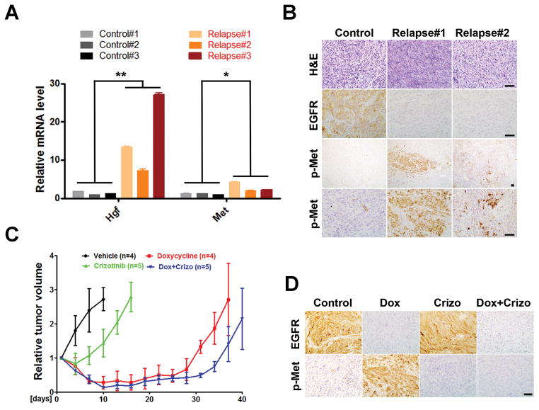
Hgf/Met signaling is activated in EGFR*-independent relapsed tumors. A, Total RNAs were prepared from untreated control and Dox-treated relapsed tumors and subjected to qPCR analysis for Hgf, Met and β-Actin. Results were normalized with β-Actin expression and shown as mean ± SD. Student’s t test was used for the comparison between untreated control and Dox-treated relapsed group (**, p = 0.034; *, p = 0.046). Data were from two independent experiments with triplicates. B, Met activated tumor cells were focally distributed in Dox-treated relapsed tumors. Shown are representative images of untreated control and relapsed tumor sections stained for H&E, EGFR and phospho-Met (p-Met). C and D, Met inhibitor had limited effect on iEIP tumor growth and relapse prevention. Mice with subcutaneously grafted iEIP glioma cells were treated with vehicle (n = 4), crizotinib (n = 5), Dox (n = 4), or Dox + crizotinib (n = 5). Day 0 represents the day when treatment was initiated. Tumor growth was measured at indicated time and calculated relative to initial tumor volume. The data are presented as mean ± SD.

