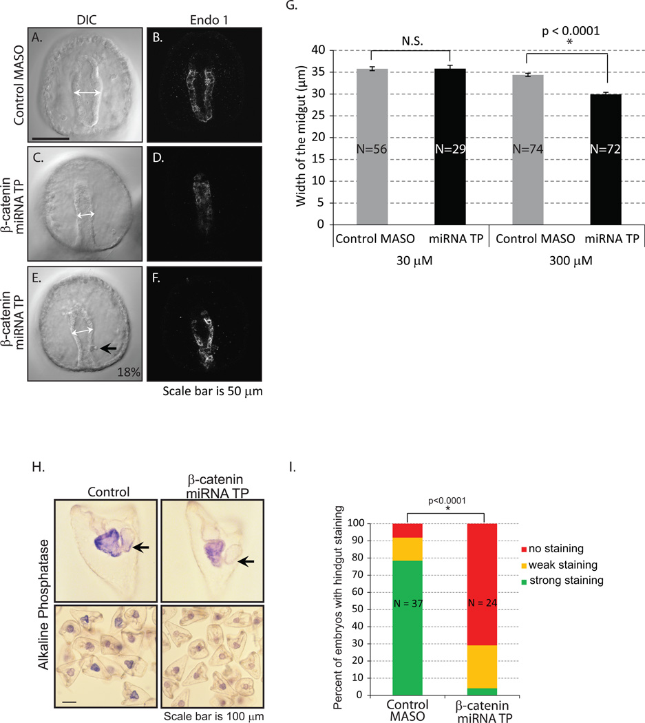
Fig. 5. Removal of miRNA regulation on β-catenin results in aberrant gut morphology.
Gastrulae were immunolabeled with Endo1, an antigen expressed in the midgut and hindgut of the embryo (Wessel et al., 1990), and imaged with an LSM 780 scanning confocal microscope. (A–B) Gastrulae injected with control MASO had a straight tubular gut that is lined with a single layer of epithelial cells. (C–F) β-catenin miRNA TP treatment resulted in embryos with narrower gut structure. (E–F) 18% of the 97 embryos miRNA TP injected embryos had an aberrant hindgut structure (black arrow) (2 out of 4 biological replicates). (G) The width of the midgut was measured with embryos injected with either 30 µM or 300 µM stock of β-catenin miRNA TP (white arrows). Only the 300 µM miRNA TP treated embryos had a significantly narrower gut than the control. *Student T-test, p<0.0001. N is the total number of embryos imaged and phenotyped. N.S. = not significant. (H) The alkaline phosphatase staining was used to assess endodermal differentiation of the larvae. Control MASO-injected embryos have more intense alkaline phosphate staining than the β-catenin miRNA TP treated embryos. Many of the embryos exposed to the β-catenin miRNA TP lack alkaline phosphatase staining in the intestine (hindgut) (arrows). (I) The percentage of embryos with hindgut staining in the miRNA TP treatment group is significantly lower than in the control embryos. *Fisher’s Exact Test (no hindgut staining vs. hindgut staining, p<0.0001). N is the total number of embryos examined.

