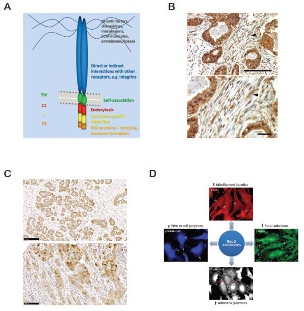
Figure 3.
A. Diagram of syndecan structure, showing some interactions and functions of the constituent domains. B. Intraductal invasive carcinoma grade III showing stromal staining (arrowheads) with mouse anti-syndecan-1 monoclonal antibody 11A9-14. Bar=100µm. C. Ductal hyperplasia (upper panel) and invasive ductal carcinoma grade III (lower panel) stained for syndecan-2 using monoclonal antibodies. Bar=100µm. D. Impact of syndecan-2 (Sdc-2) depletion on the behavior of triple negative MDA-MB231 cells. Cytoskeletal alterations include junction formation and microfilament bundle formation. Increased adhesion also results in decreased invasion and degradation of type I collagen gels.

