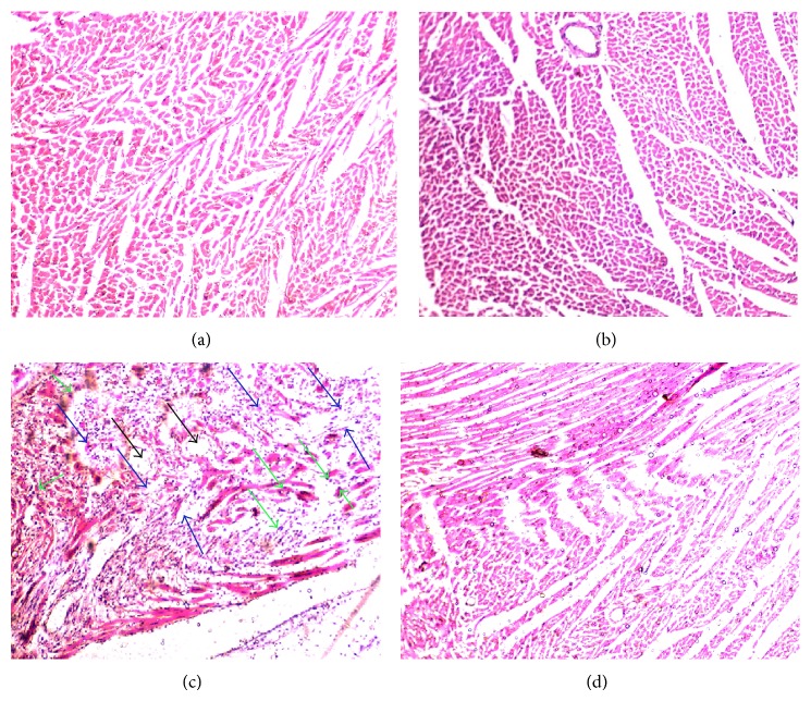
Figure 3.
Effect of TH on histopathological changes observed on the ventricular wall of heart muscle. (a) Group 1: photomicrograph of histopathological examination of stained myocardial tissue section from normal control rats showing normal cardiac muscle fibers. (b) Group 2: TH-treated (3 g/kg) heart showing normal cardiac muscle fibers with no pathological changes. (c) Group 3: ISO alone (85 mg/kg) treated heart showing separation of cardiac muscle fibers (blue arrows) and edematous intramuscular spaces (black arrows) with infiltration of inflammatory cells (green arrows). (d) Group 4: honey + ISO-treated heart, indicating that TH conferred some cardioprotection; markedly increased areas of normal muscle fibers can be observed. (Magnification: 40x).

