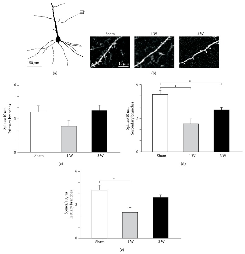
Figure 4.
Apical dendritic spine density changes in vM1 layer 5 pyramidal neurons following facial nerve axotomy. (a) Two-dimensional computer-assisted trace of layer 5 pyramidal neuron from a representative mouse sacrificed 1 week after facial nerve lesion. The small rectangle indicates the area photographed in (b). (b) Representative microphotographs of second order dendritic spines from each experimental group. (c, d, e) Quantification of layer 5 pyramidal neurons spine density in 1st, 2nd, and 3rd order apical dendrites for each experimental group. Bars and error whiskers represent the mean + SEM. 1 W, 1 week after peripheral nerve lesion; 3 W, 3 weeks after peripheral nerve lesion; ∗ P < 0.05.

