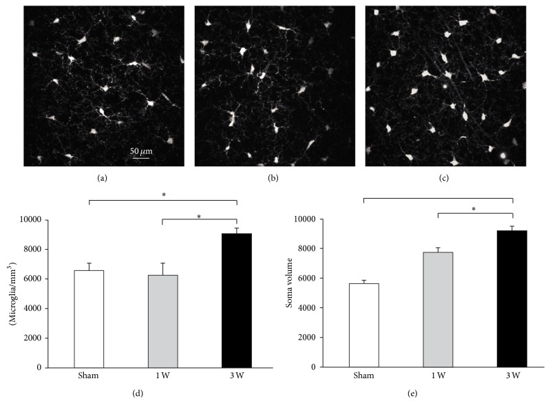
Figure 6.
Increased microglial cell density and soma area surrounding vM1 layer 5 pyramidal neurons following facial nerve axotomy. (a, b, c) Microphotographs of microglia cells around vM1 layer 5 pyramidal neurons from representative sham (a), 1 week (b), and 3 weeks (c) mice. Quantification of cell density (d) and soma area (e) for microglial cells surrounding vM1 layer 5 pyramidal neurons from each experimental group. Bars and error whiskers represent the mean + SEM. 1 W, 1 week after peripheral nerve lesion; 3 W, 3 weeks after peripheral nerve lesion; ∗ P < 0.05.

