Fig. 1.
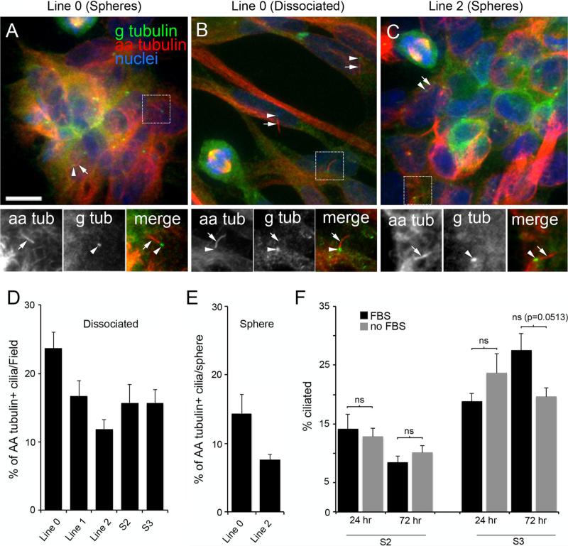
Subsets of cultured GBM cell lines derived from human GBM specimens stain positively for primary cilia. (a–c) Sphere aggregates and dissociated Line 0 (a, b) and Line 2 (c) cells were immunostained for gamma-tubulin (G-tubulin or g tub) (green) and acetylated alpha-tubulin (AA-tubulin or aa tub) (red). G-tubulin is enriched in basal bodies and spindle poles, and AA-tubulin is enriched in spindle fibers and the cilia axoneme but can also be detected throughout the cell. The spatial relationship of the staining of these two proteins was used to identify cilia. All images are maximum projections from confocal z-stacks. Scale bar (a) = 10 lm. In a–c, magnified images of the boxed regions showing the staining patterns for aa tub (arrows) and g tub (arrowheads) are displayed below the larger image. The adjacent localization of g tub and aa tub is clearly evident in the merged images. Additional cilia are indicated in the larger images using arrows/arrowheads. d Percentage (±SEM) of AA-tubulin + ciliated cells/field for the indicated cell lines, 24 h after seeding onto glass coverslips. e Percentage (±SEM) of AA-tubulin + ciliated cells/sphere for indicated lines, 2 h after seeding onto glass coverslips. f Percentages (±SEM) of S2 or S3 cells that were ciliated at either 24 or 72 h after serum removal compared to control cells maintained in FBS. Groups were compared using a 2-way ANOVA followed by Fisher's PLSD post hoc analysis. ns not significant

