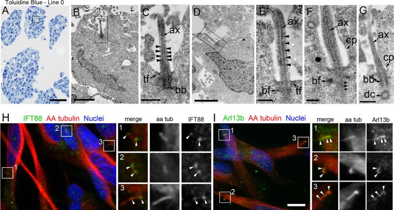
Fig. 2.
Ultrastructure of Line 0 cilia and detection of IFT88 and Arl13b along their axonemes. a Semithin sections of Line 0 spheres stained with toluidine blue. The box approximates regions where EM analysis was performed. b Low magnification of a cilium (dotted box) extending out of a cell. c Higher magnification of the cilium in (b) reveals a docked basal body anchored by transition fibers and an axoneme comprised of organized, parallel arrangements of microtubules (arrowheads). d Another example of a cilium (dotted box) extending out of the cell body, whose axoneme contained organized, parallel arrangements of microtubules (e, arrowheads). f, g Additional high magnification images of cilia showing axonemes extending out of the ciliary pocket. Typical parallel arrangements of microtubules that impinged on the basal foot (f, arrowheads) and daughter centrioles arranged perpendicularly to basal bodies were observed (g). h Immunostaining for IFT88 (green) revealed localization to the bases, axonemes, and tips of Line 0 cell cilia. Boxes 1–3 highlight IFT88 + cilia that are shown in the magnified images on the right (arrowheads). i Immunostaining for Arl13b (green) revealed localization along the ciliary axonemes and tips. Boxes 1-3 highlight Arl13b + cilia that are shown in the magnified images on the right (arrowheads).ax axoneme, tf transition fibers, bb basal body, bf basal foot, dc daughter centriole, cp ciliary pocket. Scale bars in A = 100 μm, B, D = 2 μm, C = 500 nm, E = 250 nm, F = 200 nm, G = 500 nm, I = 10 μm

