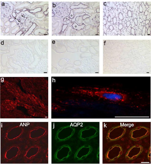
Figure 3. ANP production and binding sites in the kidney and heart.

(a – c) ANP mRNA expression obtained by in situ hybridization. (a) In the renal cortex, ANP mRNA labeling is shown in podocytes (arrowheads) and distal tubules. (b) Typical formation of connecting tubules (circles) around an artery within a cortical region is presented. (c) Medullary collecting ducts and interstitial cells labeled for ANP mRNA. (d – f) ANP mRNA sense probes were applied as a negative control showing corresponding areas of the kidney. No signal is obvious. (g and h) Immunohistochemical detection of ANP protein in the heart atrium. ANP expression in (g) low and in (h) greater detail image demonstrates the typical perinuclear vesicle staining. (i – k) Double labeling of ANP with AQP2, localization of ANP protein is exclusively present in the medullary collecting duct within the papillary tip. Magnification: calibration bar = 20 μm
