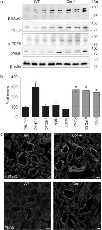
Figure 5. Alterations in the abundance of β-ENaC and ANP induced signaling components in Cor−/− mice compared to wild type mice.

(a) Representative western blots of β-ENaC and PDE5, phospho-PDE5 (p-PDE5), PKGII and β-actin (as loading control) are shown. Strongly increased abundance is found for β-ENaC, PDE5, p-PDE5 and PKGII in Cor−/− mice compared to wild type mice. (b) Corresponding densitometric evaluations of western blot analysis on ENaC subunits, Na/K-ATPase (NKA), aquaporin-2 (AQP2) and on PDE5, p-PDE5 and PKGII. Values are means ± SE; n=5 per group; *P < 0.05. (c) Immunohistochemical labeling of kidneys from Cor−/− mice show increased signal intensity of β-ENaC and PKGII in medullary collecting duct cells compared to wild type mice. Magnification: calibration bar = 20 µm
