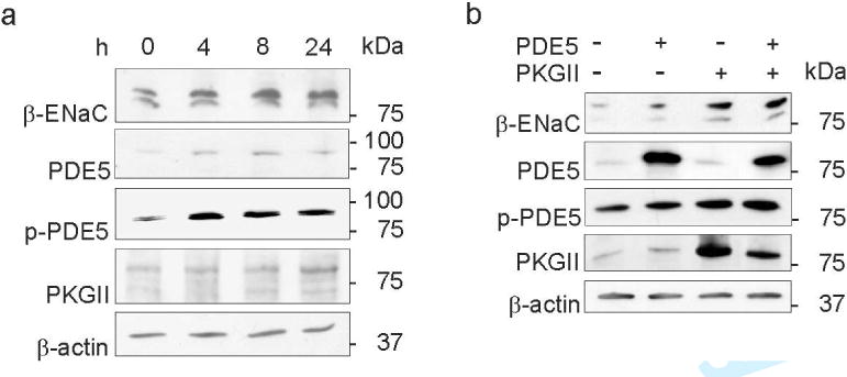
Figure 6. Alterations in expression levels of β-ENaC and ANP signaling components after cGMP reduction and PDE5 and PKGII overexpression in mpkCCDcl4 cells.

(a) Western blots of β-ENaC, PDE5, phospho-PDE5 (p-PDE5), PKGII and β-actin (as loading control) on mpkCCDcl4 cells underwent reduction in cGMP for 0, 4, 8 and 24 hours (h) are presented. Increases in β-ENaC, PDE5, p-PDE5 and PKGII abundance are shown. (b) Western blots of β-ENaC, PDE5, p-PDE5, PKGII and β-actin (as loading control) on transiently transfected mpkCCDcl4 cells using pcDNA3.1 (mock), PDE5 and PKGII constructs. Transient transfection of PDE5 and PKGII results in augmented β-ENaC abundance and transfection of PKGII phosphorylates PDE5. Values are means ± SE; n=8 per experiment; *P < 0.05.
