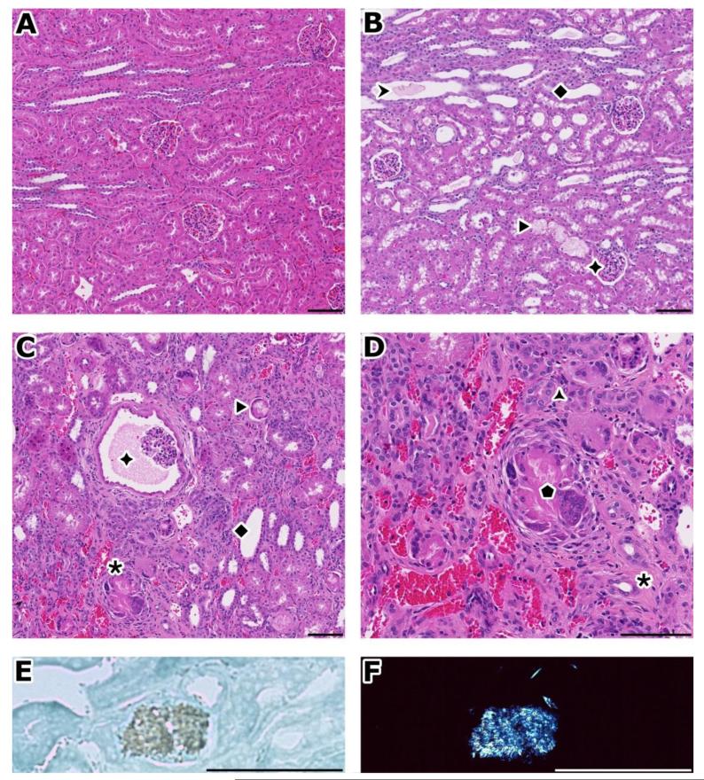
Figure 3.
Pathologic findings of toxicity study in rabbits
A) normal kidney. B) after 7 days, accumulation of granular material (➤) or spicules (▶) in dilated and basophilic corticotubules and collecting ducts (◆). Glomerules are normal (✦). C) after 1 month, there is extensive cortical damage with chronic tubulo-interstitial inflammation (✱), see prominent Bowman’s space dilation (✦). D) Close up of inflammation from figure C); regenerating tubules (basophilic) with thick basement membrane ( ), diffuse infiltrate (mainly mononuclear), congestion, interstitial fibrosis (✱), giant cells around acicular clefts (
 ). E) Appearance of an acicular crystal under bright field illumination. F) same birefringent crystal than in E), under polarization. This foreign material deposited in tubules forms spicules as in this example or can also be granular.
A-C: H&E, original magnification: 10×. D: H&E, original magnification: 20×. E, F: frozen section, methylene blue, original magnification: 40×. Bar: 100 μm

