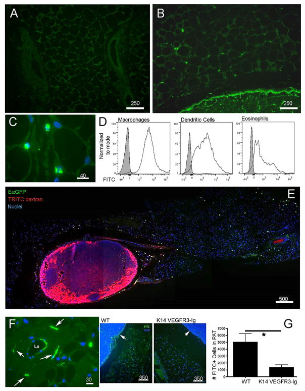
Figure 3. Passage of soluble antigens from collecting lymphatic vessels to adipose tissue phagocytes.
A, B) Low power cross-sections depicting PAT outside of the brachial LN 18 h after application to the skin surface of a contact sensitizer containing FITC (green) as a hapten (Panel B) compared to the same PAT without FITC painting in Panel A. C) Higher power view of FITC+ cells (green) in PAT as in B.D) Flow cytometric analysis to quantify FITC uptake 18 h after FITC skin painting in DCs (CD45+CD11c+MHCII+MERTK-CD64-), macrophages (CD45+MERTK+CD64+), and eosinophils (CD45+MERTK-SiglecF+). E) Low power tile-reconstructed view of a cross-section of PAT and brachial LN 20 h after EαGFP (green) was administered intradermally (i.d.) and 10 min. after 70 kDa TRITC-dextran (red) was injected i.d. F) Higher power view of PAT cross-section 10 min after FITC-dextran (green) i.d. shows a collecting vessel wall with dextran-enriched cells nearby (white arrow). G) Mice with defective lymphatics resulting from expression of VEGFR3-Ig from the K14 promoter and their littermate WT controls were subjected to FITC skin painting for 18 h. FITC+ cells in PAT cross-sections were counted (n=3 per group). Arrows point to the subscapsular sinus in each strain. Number above scale bars corresponds to number of microns. All images shown were representative of at least 3 animals per group.

