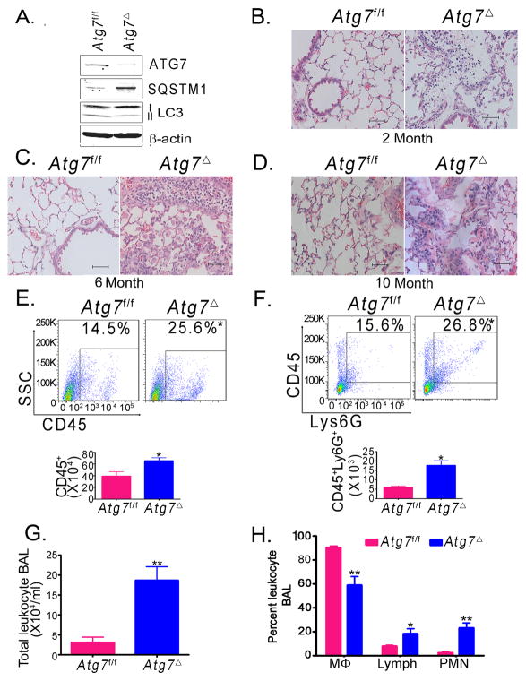
Fig. 1. Atg7Δ mice presents with spontaneous lung inflammation.
Bone marrow-derived macrophages (BMDM) from wild type (Atg7f/f) or Atg7Δ mice were analyzed by immunoblotting (A). Lung tissues from two (B), six (C) or ten (D) month old mice were analyzed by hematoxylin and eosin (H&E) staining. Flow cytometry analysis of single cell suspensions of the lungs was done using the pan leukocytic marker anti-CD45 (E) or anti-Ly6G (F). Quantification graphs are shown. Bronchoalveolar lavage (BAL) from 2–4 months old Atg7Δ or control Atg7f/f mice was analyzed for total cell count (G) or differential cell count (H). Mϕ, macrophages; Lymph, lymphocytes; PMN, polymorphonuclear neutrophils. Data are mean ± SEM, n=3 to 8 mice/genotype, *p<0.05, ** p<0.01 Scale bar, 50 μm.

