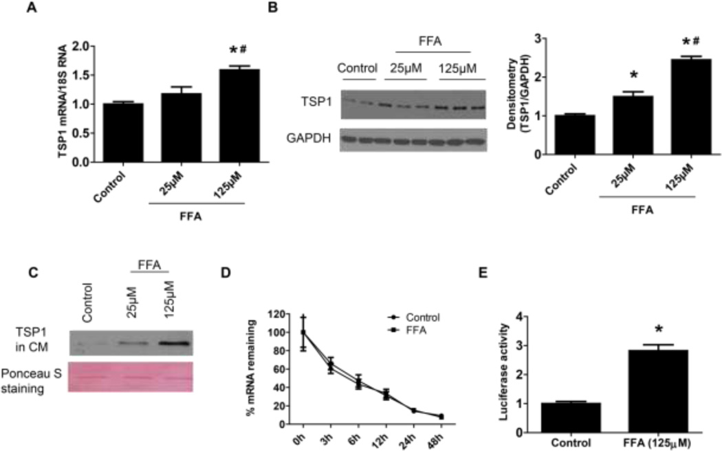
Figure 3. FFA treatment stimulated TSP1 expression in podocyte at the transcriptional level.
FFA treated human podocytes were harvested for analyzing TSP1 expression in mRNA (A) and protein (B) levels by Real-time PCR and Western blot assay, respectively. TSP1 protein levels in the conditioned media (CM) were determined by immunoblotting. The ponceau S staining was shown as loading control (C). The effect of FFA on TSP1 mRNA stability was determined by adding actinomycin D to inhibit de novo RNA synthesis after 24-h FFA treatment (D) and the effect of FFA on TSP1 transcriptional activity was tested by transfection with a mouse full-length TSP1 promoter-luciferase before 24-h FFA treatment (E). Data are presented as mean ± SE (n = 3 individual experiments). *, P <0.05 vs. control group; #, P <0.05 vs. 25 µM group.

