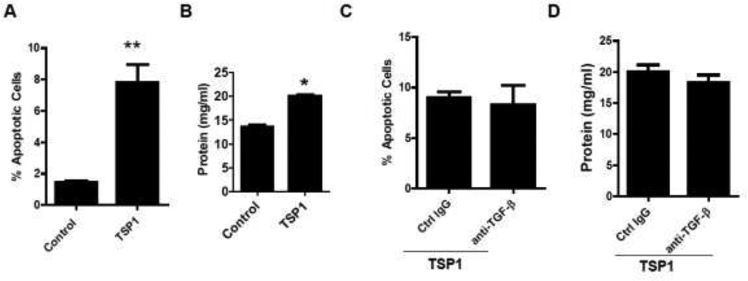
Figure 6. TSP1 treatment induced podocyte apoptosis and functional change through a TGF-β independent pathway.
Human podocytes were treated with TSP1 (1µg/ml) for 24 h. Then Annexin V/PI staining for podocytes (A) and podocyte BSA filter assay (B) were performed. Human podocytes were pretreated with anti-TGF-β 1−3 antibody (15µg/ml) or control IgG for 30 minutes and then treated with TSP1 for 24 hours. After treatment, apoptotic cells (C) and BSA filter assay (D) were analyzed. Data are presented as mean ± SE (n = 3 individual experiments). *, P <0.05 and **, P<0.01 vs. control group

