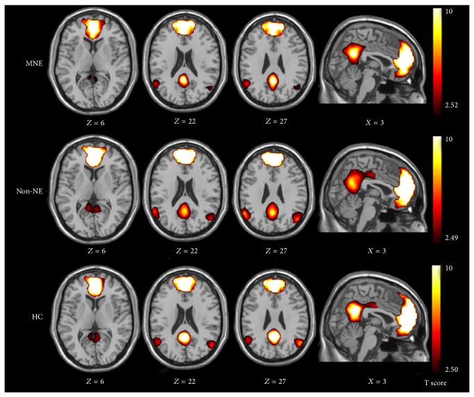
Figure 5.
Spatial maps of default mode network (DMN) in minimal nephritic encephalopathy (MNE), nonnephritic encephalopathy (non-NE), and healthy control (HC) groups. DMN of the HCs consists of bilateral posterior cingulate cortex and adjacent precuneus, angular gyri, anterior cingulate cortex, medial frontal cortex, and superior temporal cortex (P < 0.05, false discovery rate corrected). A similar DMN but with some diffuse impairment of brain areas is shown on images of MNE and non-NE patients compared with those in HCs (P < 0.05, false discovery rate corrected).

