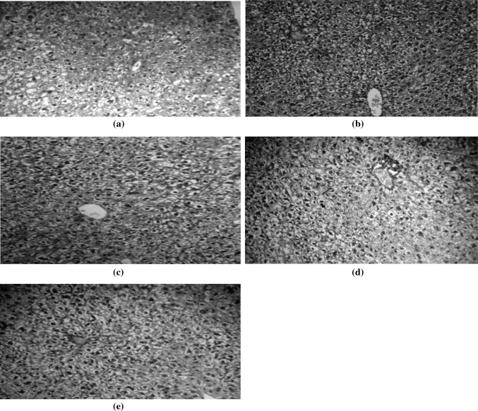
Fig. 1.
Histopathological examination of hepatic tissues. a Normal hepatic tissue sections showing normal cellular architecture with no sinusoidal growth pattern, or increased nuclear/cytoplasmic ratio (H&E × 100). b Hepatic tissue section of cancer control animals showing cellular and nuclear pleomorphism, multi-nucleated giant cells and increased nuclear/cytoplasm (N/C) ratio (H&E × 100). c Hepatic tissue section of Lawsonia-treated animals showing marked decrease in the N/C ratio, and less prominent nuclei (H&E × 100). d Hepatic tissue section of Octreotide-treated group showing marked decrease in the N/C ratio (H&E × 100). e Hepatic tissue section of (Lawsonia and octreotide)-treated animals showing marked decrease in cellular & nuclear pleomorphism and N/C ratio, and less prominent nuclei (H & E × 100)

