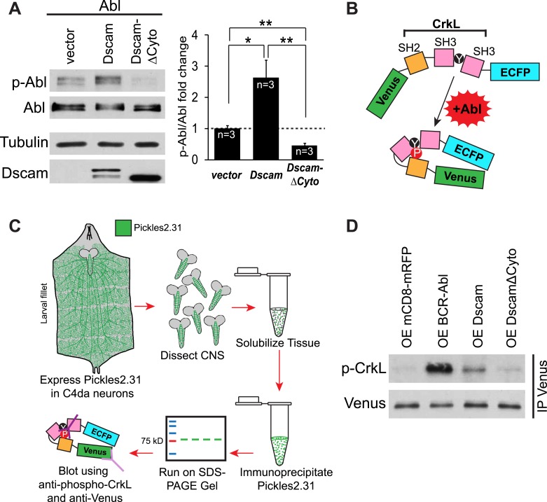
Figure 3. Dscam activates Abl kinase in culture and in vivo.
(A) Dscam activates Abl in cultured S2 cells. Abl activation was examined in S2 cell lysates transfected with indicated constructs by using anti-phospho-Y412-Abl antibody. The intensity of phospho-Abl was quantified, normalized to total Abl::Myc, and presented as bar graph (n = 3) (A, right). (B) Schematic of Pickles2.31, an Abl activity reporter that uses phosphorylation of CrkL to report Abl kinase activity. Pickles2.31 is composed of a fragment of human CrkL that contains an Abl phosphorylation site, Y207, sandwiched between ECFP and Venus. Phosphorylation of Pickles2.31 by Abl can be detected with an anti-phospho-Y207-CrkL (p-CrkL) antibody. (C) Schematic of in vivo assay for detecting Abl activity in C4da presynaptic terminals. Pickles2.31 is specifically expressed in C4da neurons. As can be appreciated from the larval fillet diagram (left), the cell bodies and dendrites of C4da neurons reside in the larval body wall while their presynaptic terminals reside in the CNS. To assay Abl activity only in presynaptic terminals, larval CNS are dissected out and solubilized into lysates. Pickles2.31 in the presynaptic terminals is then immunoprecipitated with an anti-Venus antibody (left). After running on an SDS-PAGE gel, Pickles2.31 expression level can be assayed using an anti-Venus antibody, while the phosphorylation of Y207, a proxy for Abl activity level, can be ascertained by western blotting with a p-CrkL antibody. (D) Dscam activates Abl in presynaptic terminals in vivo. Overexpression of BCR-Abl leads to a robust increase in p-CrkL staining of Pickles2.31 when compared to the mCD8-mRFP control. Similarly, overexpression of Dscam leads to consistent, though less extreme, increase in p-CrkL when compared to control. In contrast, overexpression of DscamΔCyto is indistinguishable from the mCD8-mRFP control. This is a representative blot of three experimental repeats.
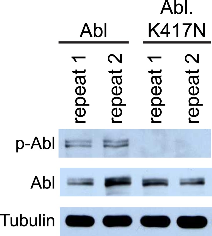
Figure 3—figure supplement 1. Phospho-Y412-Abl antibody specifically reports Abl activation.


