. 2015 May 19;4:e05196. doi: 10.7554/eLife.05196

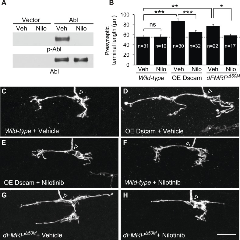
Figure 4. Pharmacological inhibition of Abl mitigates the neuronal defects caused by increased Dscam expression in vivo.

(A) Nilotinib inhibits Drosophila Abl kinase. S2 cells were transfected with either Myc-vector or Abl::Myc, and then treated with either vehicle (DMSO) or 5 μM nilotinib for 6 hr. Total lysates were subjected to western blot analysis with phospho-Y412-Abl (p-Abl) (top) and Myc antibodies (bottom). (B) Quantification of the presynaptic terminal length of the indicated genotypes and drug treatment. Sample number is shown inside each bar. (CH) Nilotinib treatment mitigates presynaptic arbor enlargement caused by Dscam overexpression (OE Dscam, D and E) and by dFMRP mutations (dFMRPΔ50M, G and H). Nilotinib treatment alone does not affect presynaptic terminal growth (F). The arrowhead in each panel points to the location where an axon elaborates the presynaptic terminal arbor. The MARCM technique was used to generate and visualize single presynaptic terminals of mutant C4da neurons. Drosophila larvae were raised in the presence of either 380 μM nilotinib or vehicle (DMSO) for 4 days before the analysis. Scale bar is 10 μm.

DOI: http://dx.doi.org/10.7554/eLife.05196.011

Figure 4.

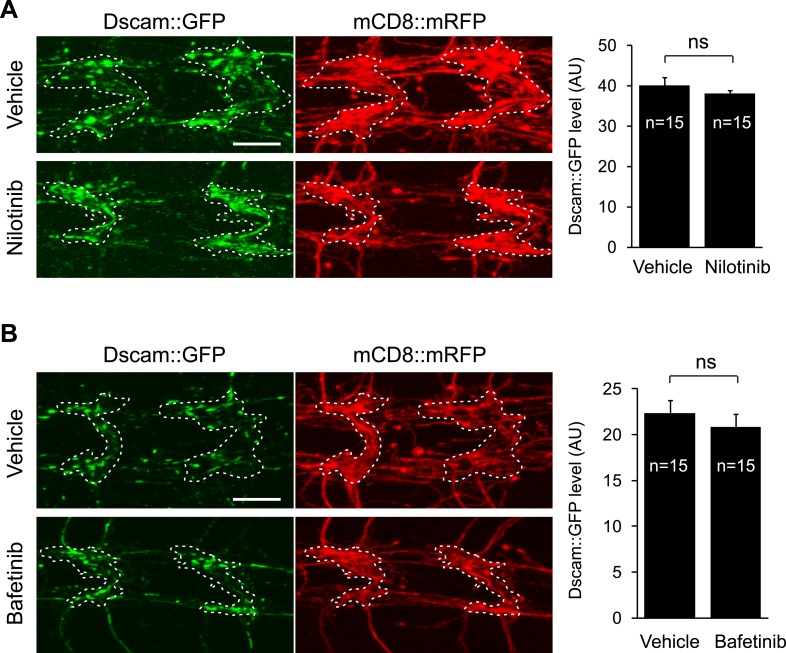
Figure 4—figure supplement 1. Nilotinib and bafatinib do not reduce Dscam transgene expression.

Figure 4—figure supplement 1.

Example images of C4da presynaptic terminals expressing Dscam::GFP in animals fed either vehicle (A and B, top), 380 µM nilotinib (A, bottom), or 125 µM bafetinib (B, bottom) throughout larval development. Images of mCD8::mRFP are shown to indicate the neuropil regions used for the quantifications (white dotted line). Scale bar is 10 μm. Quantification of the fluorescence of the Dscam::GFP transgene in neuropil region is shown on the right. Sample number is shown inside each bar.

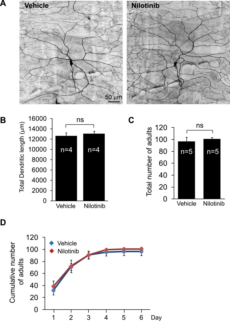
Figure 4—figure supplement 2. Nilotinib treatment does not cause defects in dendritic development or adult viability.

Figure 4—figure supplement 2.

(A and B) Nilotinib does not affect dendritic development. After egg collection, the animals were raised on food containing either vehicle (DMSO) or 380 µM nilotinib for 4 days. C4da dendrites were visualized by expressing mCD8::GFP with ppk-Gal4 (A). Total dendritic length was measured, quantified, and presented in the bar graph (B). Sample number is shown inside each bar. Scale bar is 50 µm. (C and D) Nilotinib does not affect the development of the flies. After egg collection, the animals were raised on food containing either vehicle (DMSO) or 380 µM nilotinib. Eclosed adults were counted on a daily basis. Total number and cumulative number of adults are shown in (C) and (D) respectively.

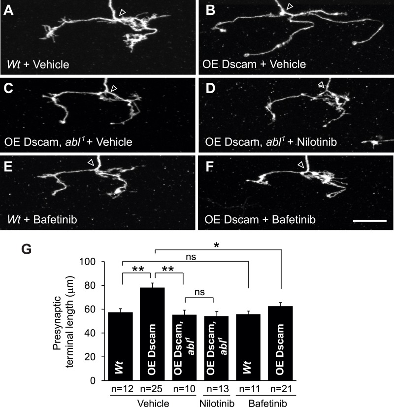
Figure 4—figure supplement 3. Nilotinib and bafetinib act through Abl inhibition to mitigate Dscam-induced presynaptic arbor enlargement in vivo.

Figure 4—figure supplement 3.

The MARCM technique was used to generate and visualize single presynaptic terminals of mutant C4da neurons. Drosophila larvae were raised in the presence of 380 µM nilotinib, 125 µM bafetinib, or vehicle (DMSO) for 4 days before the analysis. Scale bar is 10 µm. (AD) Nilotinib acts through Abl inhibition to mitigate presynaptic arbor enlargement in Dscam overexpressing neurons. Wt (wild-type, FRT2A), OE Dscam (overexpression of Dscam), OE Dscam, abl1 (overexpression of Dscam in abl1 homozygous mutations). Note that nilotinib does not further decrease the size of presynaptic arbors in abl1 neurons overexpressing Dscam(C and D). (E and F) Bafetinib mitigates presynaptic arbor enlargement in Dscam overexpressing neurons. (G) Quantification of the presynaptic terminal length of the indicated genotype and drug treatment. Sample number is shown below the x-axis.