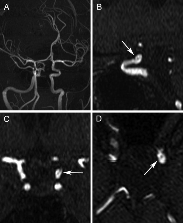
Figure 2.

MR angiography (MRA) images of the brain. The left internal carotid artery fenestration is not seen on the oblique maximum intensity projection image (A). A faint intraluminal filling defect is seen on the sagittal (B), coronal (C) and axial (D) MRA images, which can be mistaken for an intraluminal clot or focal dissection.
