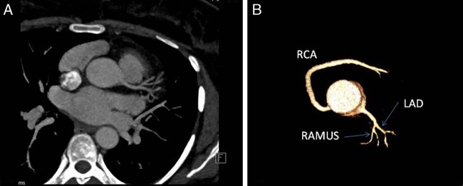
Figure 2.

Axial MIP (A) and VRT (B) images showing division of left main coronary artery into LAD and Ramus intermedius with absent LCX (LAD, left anterior descending; LCX, left circumflex artery; MIP, maximum intensity projection; RCA, right coronary artery; VRT, volume-rendering technique).
