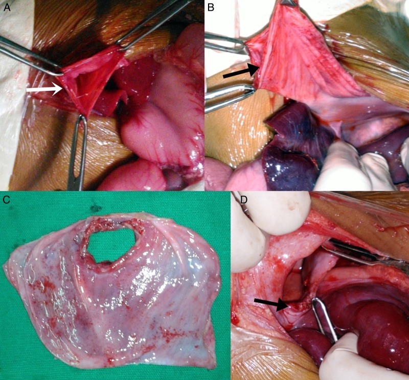
Figure 2.
Intraoperative images. (A) Defect in diaphragm (white arrow) after reducing the contents. (B) Image of the floppy left sided diaphragm, which was delivered out after reducing the content (black arrow showing site of diaphragmatic rupture). (C) Excised thinned out diaphragm with site of rupture. (D) Healthy margins (arrow) of diaphragm after excision of thin part.

