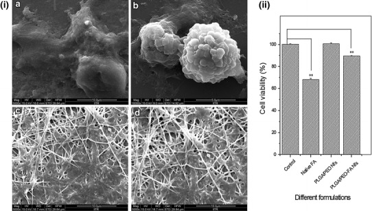
Fig. 11.

i FESEM micrographs of the HEK-293 cells incubated for 24 h on a control, b in presence of native FA, c PLGA/PEO nanofibers and d FA-encapsulated PLGA/PEO nanofibers. ii MTT assay for HEK-293 cells treated with different formulations for 24 h (A570nm). Error bars represent mean ± standard deviation for three independent experiments (n = 3). **p < 0.01 indicated statistical significant difference and (*) unmarked bar indicated statistically insignificant difference as compared to control
