
Figure 1.
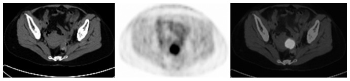
PET/CT imaging showing local recurrent rectal carcinoma in a 49-year-old female postoperative rectal cancer patient. CT scan did not find definite recurrence signs. PET imaging detected hyper-intensive radioactivity by the left side of uterus. The SUVmax was 7.5 and the T/N was 9.4. Combined PET/CT imaging indicated elevated 18F-DG uptake of the anastomosis. The reoperation confirmed a recurrent tubular adenocarcinoma of the rectum.