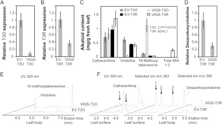
Fig. 2.
Silencing T3O (CYP71D1V2) or T3R (ADHL1) by VIGS in C. roseus leaf. Relative expression of T3O (A) and T3R (B) in C. roseus first leaf pair in comparison with the EV control plants. Internal control 60S ribosomal RNA was used for data normalization. Alkaloid contents in respective plants (C). Relative content of desacetoxyvindoline in VIGS-T3R plants (D). Biological replicates: EV-T3O, n = 5; VIGS-T3O, n = 5; EV-T3R, n = 5; and VIGS-T3R, n = 4. Error bar indicates SD. Total MIA 1–3: 3-oxidized isomers of 16-methoxytabersonine found in F (Middle). Representative LC-MS chromatograms monitored at 300 nm MIAs extracted from leaf body treated with VIGS-T3O compared with EV control (E). Representative LC-MS chromatograms of leaf surface or leaf body treated with VIGS-T3R compared with EV control (F). Isomers of 3-oxidized 16-methoxytabersonine were identified by measuring absorbance at 300 nm (Left) or by selected ion m/z= 383 (Middle). The content of desacetoxyvindoline was measured by selected ion m/z= 399 (Right). The 3-oxidized isomers of 16-methoxytabersonine could also be detected in in vitro enzyme assays with T3O-expressing yeast microsome in Fig. 3A and Fig. S2 B and F.

