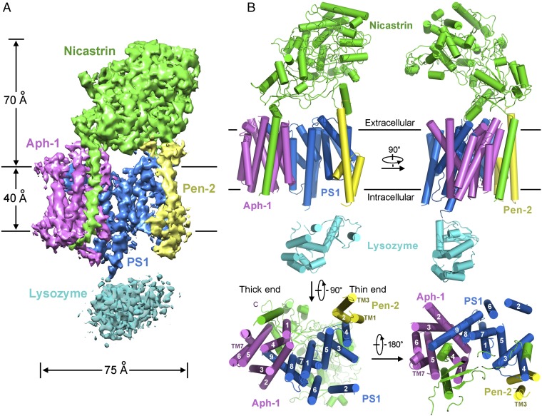
Fig. 2.
Overall structure of human γ-secretase. (A) An overall view of the EM density at 4.32-Å resolution. Densities for the four components of γ-secretase are color-coded: PS1 (blue), Pen-2 (yellow), Aph-1 (magenta), and nicastrin (green). Except TM2 of PS1, all other 19 TMs display clearly identifiable density. (B) Cartoon representation of the γ-secretase structure is shown in four perpendicular views. The four components are color-coded: PS1 (blue), Pen-2 (yellow), Aph-1 (magenta), and nicastrin (green). This coloring scheme is used in the other figures in this article. The 20 TMs assemble into a horseshoe-shaped structure. Notably, TM6 and TM7 of PS1, which harbor the two catalytic residues Asp257 and Asp385, are located on the convex side of the TM horseshoe. The structure figures were prepared using PYMOL (50).

