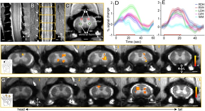
Fig. 1.
Innocuous tactile and noxious heat stimuli evoked fMRI activations in the gray matter of cervical spinal cord. (A) Middle sagittal MTC image (0.75 mm thickness) of cervical spine showed the entering roots of C4–C7 afferents (white fiber bundles). (B) Coronal MTC image (0.5 mm thickness) shows the placement of five axial imaging slices, with the center slice at the C5/C6 level. Alternating white and gray (bright) matter strips of the spinal cord were apparent. Surrounding cerebrospinal fluid was present as two white strips on both the left and right sides of the spinal cord. Nerve afferents entering roots (indicated by white arrows) were also visible and allowed identification of each segment of the cervical spinal cord. (C) MTC axial image (3 mm thickness) revealed clearly the butterfly-shaped spinal gray matter. Red circles indicate the dorsal sensory horns, and green circles show the ventral horns. Surrounding back region comprises bone structures. (D) Event-averaged time courses of fMRI signal (%) to innocuous tactile (8 Hz vibration) stimulation at four horns (LDH, left dorsal horn; LVH, left ventral horn; RDH, right dorsal horn; RVH, right ventral horn) and one white matter (WM) control. The color shadow around each color line indicated the ±SE of fMRI signal change. The red line along the x axis showed the 30-s stimulus duration. (E) Event-averaged time courses of fMRI signal (%) to noxious heat (47.5 °C) stimulation at four horns and one white matter control region. Stimulus duration is 21 s. (F) fMRI activation map to tactile stimulation of distal finger pad of D2 on left hand (thresholded at t = 4; see color scale bar in 5) on five consecutive axial MTC structural images (1–5, from tail to head). (Black scale bar in 5, 1 mm.) (G) fMRI activation map to noxious heat (47.5 °C) stimulation of distal finger pads of D2 and D3 on the right hand (thresholded at t = 3; see color scale bar in 5). D, dorsal; L, left; R, right; V, ventral.

