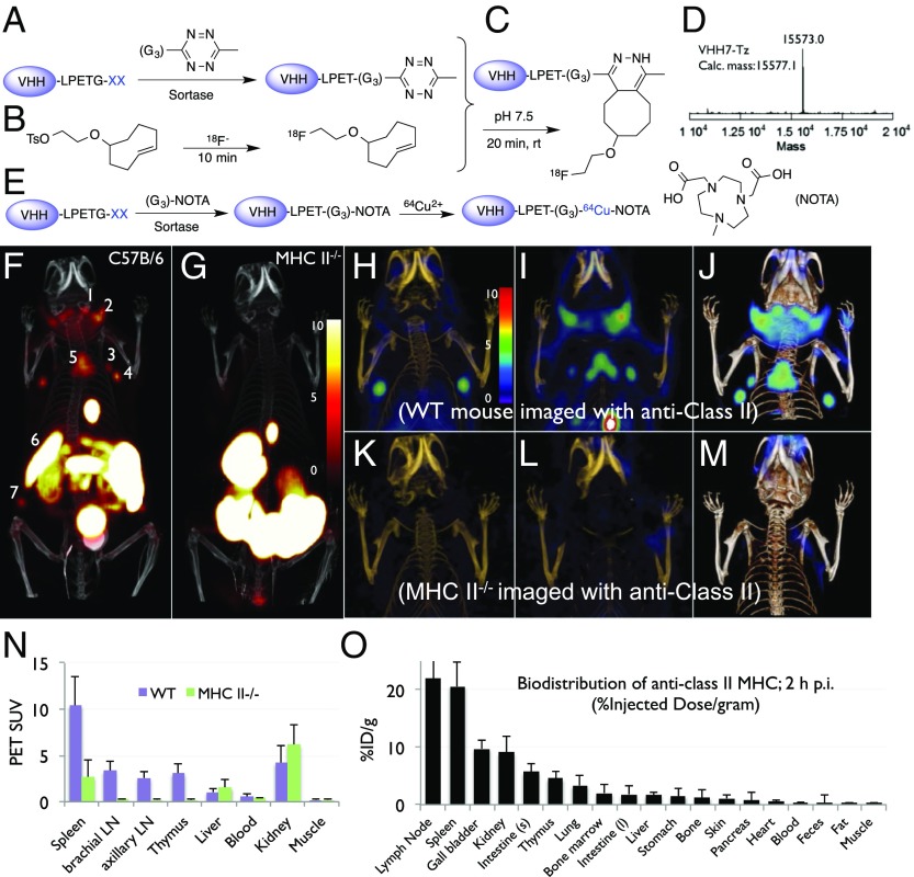
Fig. 2.
(A–E) Site-specific 18F or 64Cu labeling of single-domain antibodies (VHHs) using sortase. (A) A single-domain antibody fragment (VHH), equipped at its C terminus with the LPXTG sortase recognition motif followed by a His tag, is incubated with sortase A, which cleaves the threonine–glycine bond to yield the reactive thioacyl intermediate. Addition of a peptide with N-terminal glycine residues and a functional moiety of choice enables site-specific modification of the VHH. We thus modified a VHH with a (Gly)3-tetrazine (Tz), as confirmed by LC-MS (liquid chromatography-mass spectrometry) (D, VHH7-Tz). (B) A tosyl-TCO and 18F-K222/K2CO3 were combined to produce 18F-TCO. (C) 18F-TCO was added to the Tz-modified VHH, and, after ∼20 min, the labeled VHH was retrieved by rapid size-exclusion chromatography. (E) The sortase reaction was used to install a NOTA functionality at the C terminus of a VHH followed by addition of 64Cu2+ to produce 64Cu-VHH. (F–M) 18F-VHH7 (anti-mouse class II MHC) detects secondary lymphoid organs. (F and G) PET-CT images of class II MHC-/- (G) and C57BL/6 (F) mice 2 h postinjection of 18F-VHH7; numbers indicate (i) lymph nodes (numbers 1, 2, 3, 4, and 7), (ii) thymus (number 5), and (iii) spleen (number 6). (H and I) Coronal PET-CT images of C57BL/6 mouse imaged with 18F-VHH7, moving from anterior to posterior. In H and I, different sets of lymph nodes and thymus are visible. (K and L) Coronal PET-CT images of an MHC-II-/- mouse imaged with 18F-VHH7, moving from anterior to posterior. In neither K nor L, lymph nodes or thymus is visible. (J and M) PET-CT as maximum intensity projections of all slices for a C57BL/6 and a class II MHC-/- mouse 2 h postinjection of 18F-VHH7. In green, accumulation of 18F-VHH7 in lymph nodes and thymus. (PET scale bars have arbitrary units.) See Movies S1A, S1B, S2A, and S2B for 3D visualization of secondary lymphoid organs. (N and O) PET signals in vivo and postmortem biodistribution in all organs.

