Fig. 5.
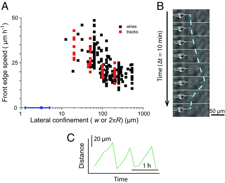
(A) Front velocity as a function of lateral confinement. Confinement is either the width w of the tracks (red points) or the perimeter 2πR of the wires (black points). Confinement controls the velocity. The blue point shows the arrest of migration on submicron wires. (B) The migration stops at very small radii, at which point the leading cell sends out periodic protrusions. The line is a guide for the eye. (C) Position of the extremity of the protrusion as a function of time. Note the asymmetric shape of these protrusion−retraction cycles.

