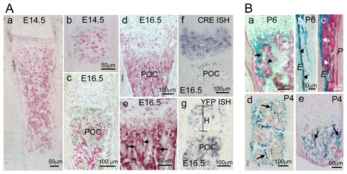
Fig. 1. Lineage tracing reveals a progeny trabecular osteoblasts derived from Col10a1 expressing cells.
(A) Anti YFP staining of Col10Cre;YFP+ bones at E14.5, before bone marrow formation, is restricted to hypertrophic chondrocytes; (a) tibia, (b) digit; (c–e). At E16.5 YFP+ cells are visible in the spongiosa; (c) E16.5 ulna, (d,e) E16.5 tibia. (f) In situ hybridization shows that Cre is expressed exclusively in hypertrophic chondrocytes (see also supplementary material Fig. S1). (g) YFP is expressed both in hypertrophic chondrocytes, ‘H’, and in cells of the spongiosa; (f,g) E16.5 humerus. POC = Primary ossification center. (B) X-gal stained sections of Col10Cre;R26R mice (a) P6 radius; (b,c) P6 humerus; (d) P4 vertebra and (e) P4 rib reveals β-gal activity in osteoblast-like cells (blue) lining trabecular bone spicules (arrows in a,d,e), in endosteal cells (E in b,c), and in osteocytes (arrows in b,c). (c) Cells in the periosteum (P) do not stain for β-gal. Counterstaining of the extracellular matrix with anti Col1 (red).

