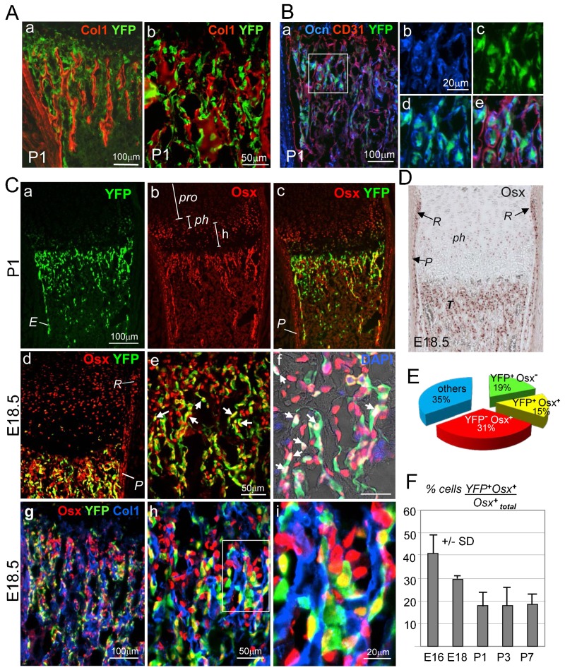
Fig. 2. Dual origin of trabecular osteoblasts in the spongiosa of Col10CreYFP+ specimens.
(A) Co-immunostaining for YFP and Col1 of a P1 Col10CreYFP+ tibia shows that YFP+ cells in the spongiosa are to a large part associated with Col1+ trabeculae. Panels a and b show different magnifications. (B) Triple staining of a Col10CreYFP+ tibia (P1) for YFP (green), osteocalcin (Ocn, blue and CD31 (red), confirms the presence of YFP+Ocn+ osteoblasts in the spongiosa, whereas endothelial cells are YFP negative. (b–e) Enlarged images of the boxed area in panel a showing single channels (b,c), two channels (d) and all three channels (e). (Ca–f) Immunofluorescence staining for Osx (red) and YFP (green) shows that a substantial number of Osx+ cells in the spongiosa and endosteum ‘E’ of P1 (a–c) and E18.5 (d–i) tibiae are YFP+ (yellow cells in c–e; white cells in f). In the periosteum ‘P’ in, all Osx+ cells are negative for YFP (c,d). pro = proliferating zone, ph = prehypertrophic zone; h = hypertrophic zone. (g–i) Triple immunostaining for Osx, Col1 and YFP: most YFP+Osx+ cells (yellow) and YFP−Osx+ cells (red) are associated with trabeculae, stained for Col1 (blue). (D) Immunostaining of paraffin sections shows Osx+ cells in the groove of Ranvier ‘R’, periosteum ‘P’, pre-hypertrophic chondrocytes ‘ph’ and trabecular bone ‘T’. E18.5 Col10CreYFP+ tibia. (E) Pie chart showing the relative amounts of chondrocyte-derived (YFP+) cells of all DAPI+ cells and other cells in the spongiosa of P2 tibiae. 15% of all DAPI+ cells are YFP+Osx+; 19% are YFP+Osx− cells and thus not osteogenic; 31% are Osx+YFP−. Cells were counted in the trabecular zone in five humeri or tibia sections (100–300 cells per field). (F) Bar graph showing ratios of YFP+Osx+ double positive (yellow) cells to total Osx+ cells (red and yellow) in the spongiosa at various stages of development. Error bars: SD.

