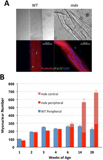
Figure 7.

Increase with age in the proportion of ‘central nuclei’ and of fibre branching in mdx mice. (A) Interference contrast and fluorescent micrographs of single fibres isolated from 1-year-old mdx and WT mice and immunostained for Pax7. These not only bear readily identifiable satellite cells (green) but also illustrate the extensive branching of fibres in older mdx but not in WT mice together with the tendency for myonuclei to be arranged in linear ‘central strings’. (B) Counts of myonuclei classified into peripheral location and ‘central’ location, the latter being classified on the basis of their distribution in linear arrays. These ‘central’ myonuclei are first seen in 6-week-old mdx mice. At 14 and 28 weeks, however, it is notable that the numbers of peripherally located myonuclei per fibre are not substantially different from those of the WT and that the centrally located nuclei account for all of the excess myonuclei found in older mdx muscles.
