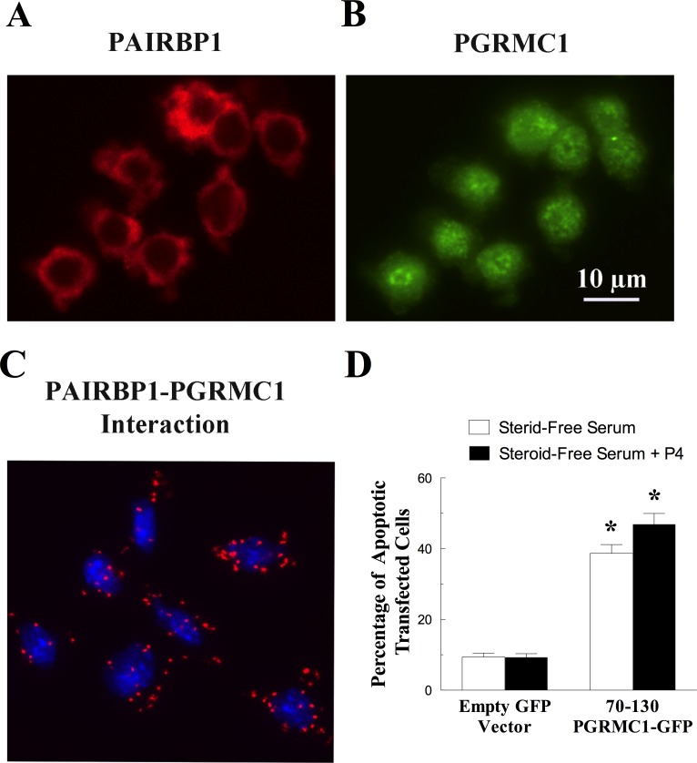
FIG. 6.
The localization and interaction of PAIRBP1 and PGRMC1 in granulosa cells maintained in serum-supplemented medium. The localizations of PAIRBP1 shown in red (A) and PGRMC1 shown in green (B) were determined by immunocytochemistry, and the interaction between PAIRBP1 and PGRMC1 was revealed by PLA (red dots, C). In the PLA image, the nuclei were stained with DAPI (blue). The graph in D shows the effect of transfecting the PGRMC1 peptide mimic (70-130-PGRMC1-GFP) on the rate of apoptosis in granulosa cells cultured in the steroid-free serum with or without P4. In this graph, the values are expressed as means ± one standard error, with * indicating a value that is greater than (P < 0.05) granulosa cells transfected with empty vector.

