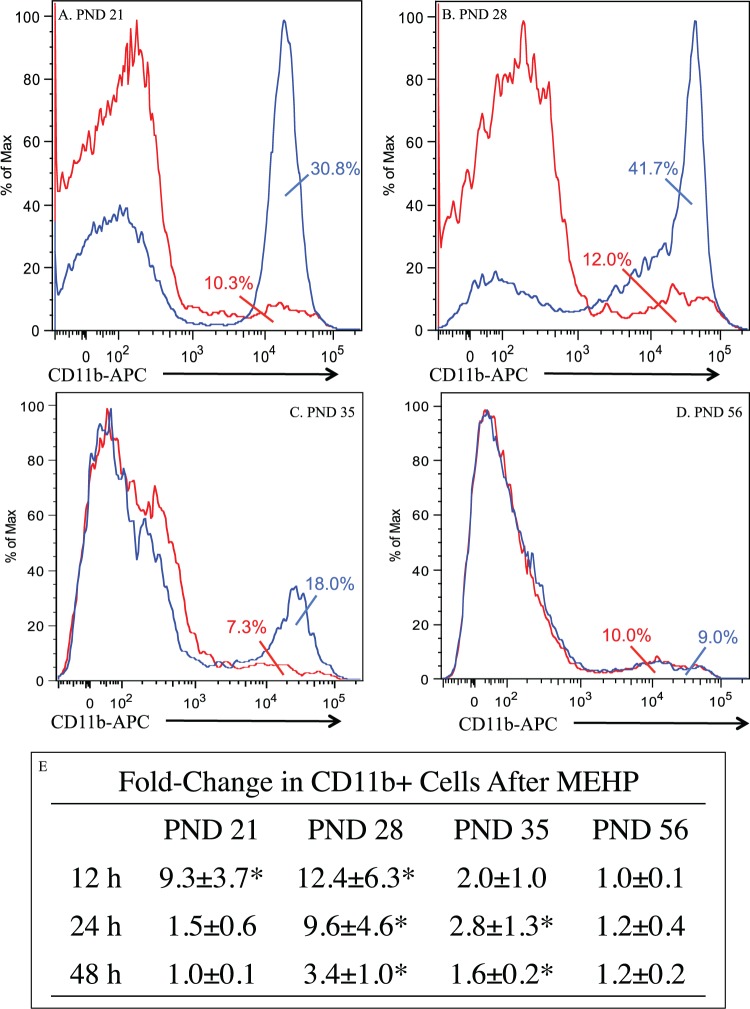
FIG. 1.
Age-dependent MEHP-induced testicular infiltration of CD11b+ cells in rats. Expression of CD11b+ cells in single cell suspension of live testicular interstitial cells after MEHP treatment of PND 21 (A), 28 (B), 35 (C), and 56 (D) rats after 12 h of MEHP exposure. Blue represent MEHP-treated rats (1 g/kg, p.o.) and red is vehicle-treated rats (corn oil, equivalent volume). The fold-change increases in CD11b+ cells after MEHP treatment (1 g/kg, p.o.) at each age and time point are summarized in the table (E). Asterisks (*) indicate significant differences between treatments at specified time points (P < 0.10, Tukey honestly significant difference [HSD] test; PND 21: n = 4, PND 28: n = 6, PND 35: n = 6, PND 56: n = 3 per time point/treatment).

