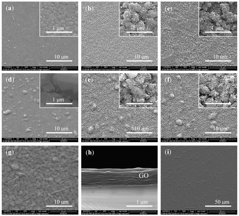
Figure 3.
Changes in the surface morphology of the electrode as observed using SEM: (a–c) non-heat-treated electrodes and (d–f) electrodes heat-treated at 320 °C. (The magnification of the images in (a–g) is × 5 K, that for images in the insets of (a–f) and (h) is × 50 K, and that for the image in (i) is × 2 K. Images of the Ag and Ag/AgCl thin films after the following steps are shown: (a) before the heat treatment, (d) after the heat treatment, (b,e) after chlorination with 50 mM FeCl3, and (c,f) after overnight storage in a saturated AgCl solution. (g) GO layer on a ready-to-use electrode. (h) and (i) show cross-sectional and top views of the pristine GO layer, respectively.

