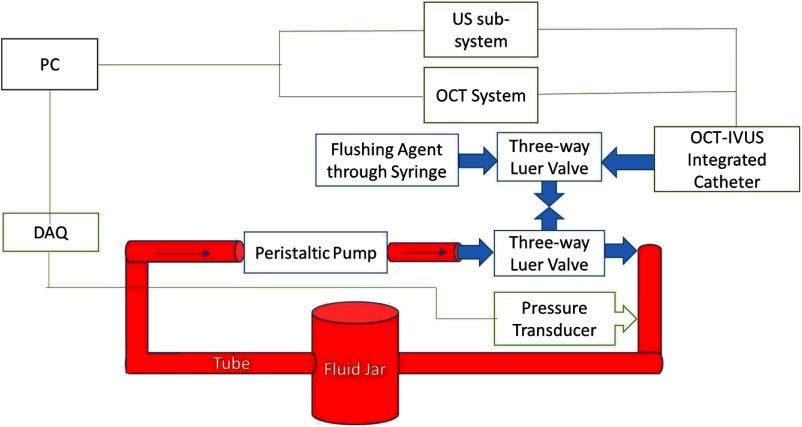
Fig. 1.
Schematic of the in vitro circulatory system model with signal acquisition devices. The personal computer (PC) acquired the optical coherence tomography (OCT) signal, intravascular ultrasound (IVUS) signal, and fluid pressure signal. Three-way luer valves were used to provide access to the closed-loop tube system and allow the imaging probe and chemicals to enter. Red areas denote where blood circulates.

