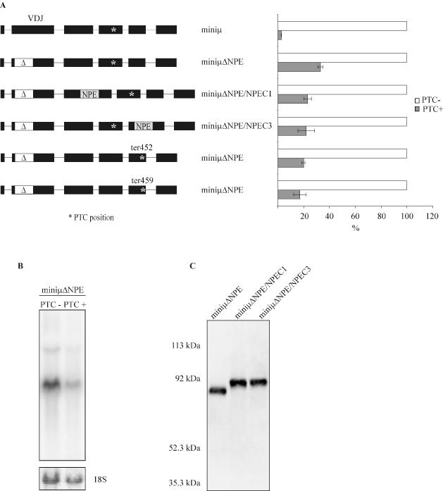
Figure 5.

The NMD-promoting element is necessary for efficient NMD of Ig-μ minigene mRNA and functions in a position-dependent manner. (A) Relative Ig-μ mRNA levels of HeLa cells stably expressing the constructs schematically represented on the left were determined by real-time RT–PCR and are shown in the right panel. For all constructs, the PTC− mRNA level (white bars) was set as 100% and the PTC+ (ter310) mRNA level (gray bars) was calculated relative to it. The Ig-μ values were normalized to endogenous GAPDH mRNA. Average values of three real-time PCR runs of one representative cell pool are shown, and error bars indicate standard deviations. The deletion in all miniμΔNPE constructs, indicated by the white region marked with Δ, is identical to the deletion in hyb8 (Figure 3) and comprises 177 bp from amino acid positions 19–76. MiniμΔNPE/NPEC1 and miniμΔNPE/NPEC3 were generated by insertion of the NPE into exon C1 or into exon C3 of the PTC− and the PTC+ version of miniμΔNPE, respectively. (B) Northern blot analysis of 15 μg RNA from the PTC− and PTC+ miniμΔNPE expressing cells analyzed in (A). As a loading control, the 18S rRNA band from the ethidium bromide-stained gel before blotting is shown in the lower panel. (C) Detection of the Ig-μ polypeptide in lysate of cells expressing the PTC− version of the indicated constructs by western blotting confirms the intactness of the respective ORFs in these mRNAs. MiniμΔNPE/NPEC1 and miniμΔNPE/NPEC3 encode for a 593 amino acids long polypeptide, the polypeptide encoded by miniμΔNPE is 59 amino acids shorter.
