
Figure 2.
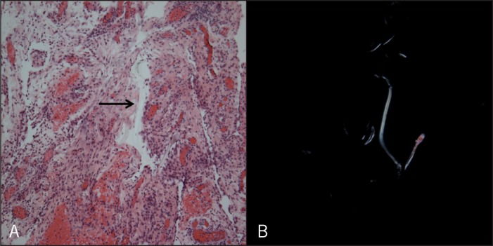
Histopathologic findings on examination of mesenteric biopsy specimens (40×). (A) Hematoxylin and eosin stain showing papillary mesothelial hyperplasia with the presence of a refractile foreign body (arrow). (B) The foreign body material is clearly illustrated when viewed under polarized light.