Figure 2.

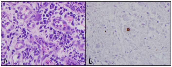
Biopsy of the mass showing (A) cytomegalovirus inclusions in HE stain and (B) immunohistochemical staining for CMV, showing positive cell with ‘owl's eye’ appearance.

Biopsy of the mass showing (A) cytomegalovirus inclusions in HE stain and (B) immunohistochemical staining for CMV, showing positive cell with ‘owl's eye’ appearance.