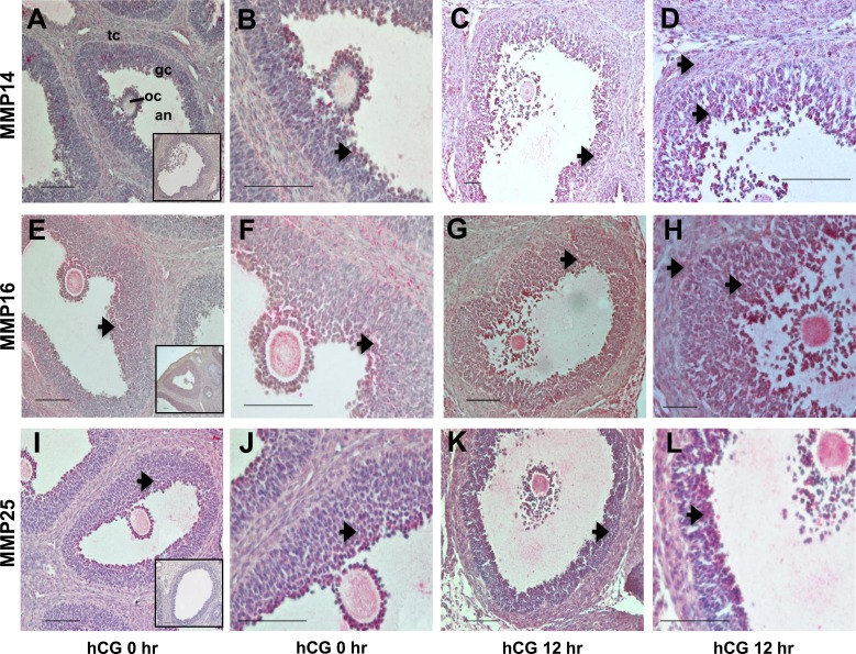
FIG. 5.
Immunohistochemical detection of MMP14, MMP16, and MMP25 in the rat ovary during the periovulatory period. Ovaries collected from eCG-primed rats at different times after administration of an ovulatory dose of hCG were processed for immunolocalization of MMP14 (A–D), MMP16 (E–H), and MMP25 (I–L). In ovaries collected at 0 h (A, B, E, F, I, and J) and 12 h (C, D, G, H, K, and L) h after hCG administration, immunoreactive MMP14, MMP16, and MMP25 protein are identified as a red reaction product (arrows). B, D, F, H, J, and L are higher-magnification views (×40) of the images in A, C, E, G, I, and K (×20), respectively. Insets in A, E, and I are ovary sections in which the primary antibodies against MMP14, MMP16, and MMP25, respectively, were omitted. The full preovulatory (0 h) or periovulatory (12 h) ovarian follicle is shown (gc, granulosa cell layer; tc, theca cell layer; an, follicular antrum; oc, oocyte). Representative photomicrographs are shown for each time point. Bar = 50 μm (×20 panels) and 100 μm (×40 panels).

