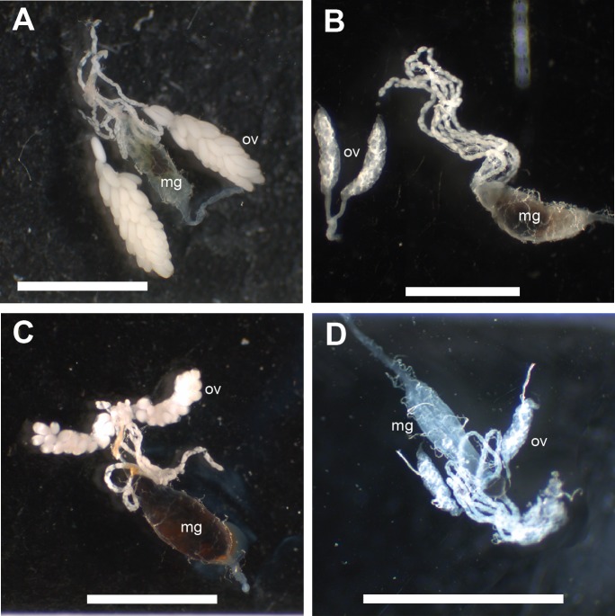
Figure 4. The effect of whole blood, RBCs and Hb meals on ovary development 72 h after feeding.
Midguts with attached Malpighian tubules and ovaries were dissected: mg, midgut; ov, ovary. Scale bars = 2 mm. (A) Fully developed ovaries from a female given whole blood (control), (B) No ovary development in females fed with PBS-buffered Hb diet, (C) Partial ovary development seen in a few females given RBC as sole protein source, (D) Unfed control female.

