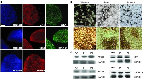
Figure 2.

Establish patient-specific cell lines. (a) Representative immunofluorescence image of pluripotency markers in established patient-specific iPS cell line. Scale bar: 100 μm. (b) Light microscopy images of cultured human iPS-RPE cells from a wild-type control donor (left), P1 (middle), and P2 (right). For upper panel, scale bar: 50 μm; lower panel, scale bar: 100 μm. (c) Immunoblot analysis of mature RPE specific marker RPE65 (65 kDa), BESTROPHIN-1 (68 kDa), MITF (59kDa), and VINCULIN (124 kDa) in iPS-RPE cells from wild-type control donor (WT), P1, and P2. GAPDH serves as the loading control.
