Figure 3.
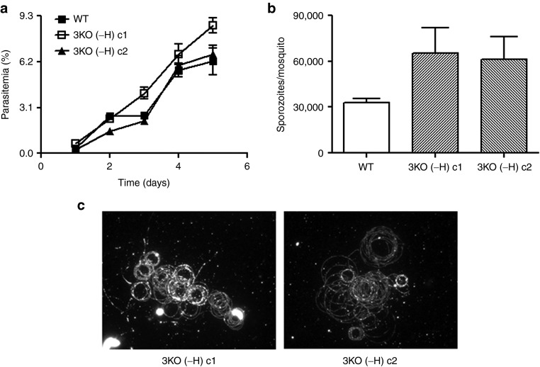
Phenotypic analysis of Pf p52−/p36−/sap1−3KO (-H) clones. (a) Comparison of asexual blood stage growth rates between wild type (WT) and 3KO (-H) clones. Cultures were initiated at 0.5% parasitemia and analyzed daily until day 5. Growth assays were performed in triplicate for each line and parasitemia plotted as mean ± SEM. Mann–Whitney U-test was used for statistical analysis. (b) Comparison of average number of sporozoites (plotted as mean ± SEM) per mosquito between WT and 3KO (-H) clones 14–16 days postfeeding of mosquitoes. Sporozoite numbers were determined three independent times in at least duplicate for each line. Mann–Whitney U-test was used for statistical analysis. (c) Staining of CS trails using Alexa Fluor 488-conjugated anti-PfCSP 2A10 antibody in motility assays of salivary gland sporozoites of WT and 3KO (-H) clones.

