
Figure 2. Adamts5 null mice have enlarged and malformed pulmonary valves.
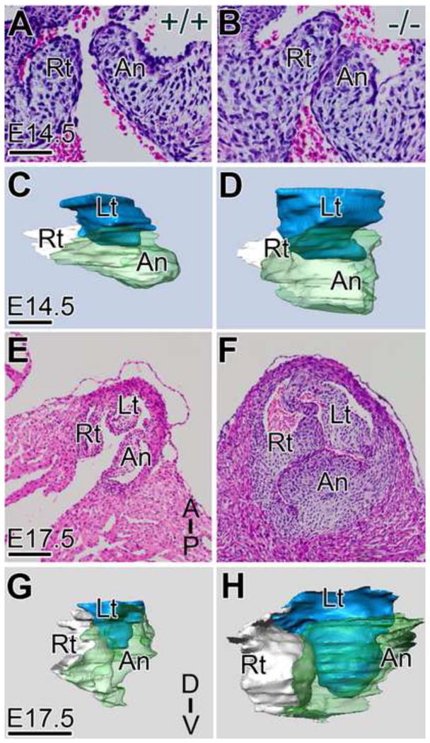
H & E stained frontal sections of the PV from WT (A, E) and Adamts5−/− (B, F) hearts at E14.5 (A, B) and E17.5 (E, F) same magnification are shown. Three-dimensional reconstructions of WT (C, G) and Adamts5−/− mice (D, H) were generated from histological sections of E14.5 and E17.5 PV. Lt- left cusp of the PV (blue); Rt- right cusp of the PV (white); An-anterior cusp of the PV (green); D-dorsal; V-ventral; Scale bars: A = 50 μm; C=75 μm; E =150 μm; G = 200 μm. Corresponding quantitative data from reconstructions are listed in Supplemental Table I.