Figure 3.
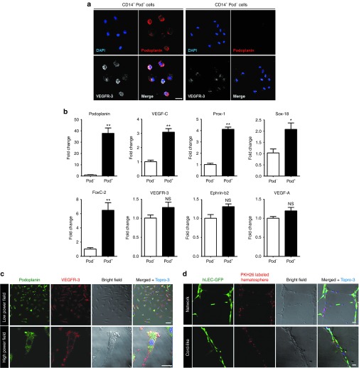
Podoplanin-positive/VEGFR-3high myeloid cells within hematospheres express lymphangiogenic characteristics. (a) Flow cytometry–sorted CD14+ Pod+ cells and CD14+ Pod− cells from day 5 hematospheres were subjected to immunofluorescence staining for podoplanin and VEGFR-3. Scale bar = 20 µm. (b) Quantitative RT-PCR of flow cytometry–sorted CD14+ Pod+ cells and CD14+ Pod− cells. Bar graphs represented the relative quantity of the lymphangiogenesis-related gene expression in the pod+ cells compared to pod− cells (*P < 0.05, **P < 0.01; n = 3 per experiment). (c) Dissociated single cells from hematospheres were seeded on 1.5% gelatin-coated dish and cultured for 24 hours. Attached cells at 24 hours were subjected to immunofluorescence staining for podoplanin and VEGFR-3. Scale bar = 50 µm. (d) Coculture of red PKH26-labeled dissociated single cells from hematospheres with lenti-GFP-transduced hLEC on Matrigel. Scale bar = 50 µm. hLEC, human lymphatic endothelial cell; NS, nonsignificant; VEGF, vascular endothelial growth factor; VEGFR, vascular endothelial growth factor receptor.

