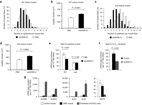
Figure 3.

The effect of myostatin/ActRIIB signaling on vascularization. All investigations were done after a 4-month treatment of wild-type and mdx mice with either soluble activin IIB receptor (sActRIIB-Fc) or PBS (controls). (a–d) Capillarization of wild-type soleus (n = 430 fibers from PBS-treated muscles (n = 3) and n = 300 fibers from sActRIIB-Fc-treated muscles (n = 3)) and mdx soleus muscle (n = 605 fibers from PBS-treated muscles (n = 3) and n = 836 fibers from sActRIIB-Fc-treated muscles (n = 4)). Histograms in (a) and (c) depict the distribution of capillaries per muscle fiber (in (%)), whereas diagrams in (b) and (d) depict the capillary domain, the fiber area per capillary (in (µm2)). Values are depicted as means ± SEM. (e) Vegf-A relative mRNA copy numbers as expressed per 106 × 18S rRNA copies in wild-type TA muscle (n = 5 for each condition). (f) Vegf-A relative mRNA-copy numbers in C2C12 myotubes following 24 hours treatment with sActRIIB-Fc in comparison to control cultures (n = 3 for each condition). (g) Relative mRNA-copy numbers of MSTN and myostatin receptors ActRIIA/B and ALK4/5 in cultures of human umbilical vein endothelial cells (HUVEC) in comparison to muscle samples from a healthy control and a patient with Duchenne muscular dystrophy. Values are shown as means ± SEM. P values were calculated using the nonparametric U-test.
