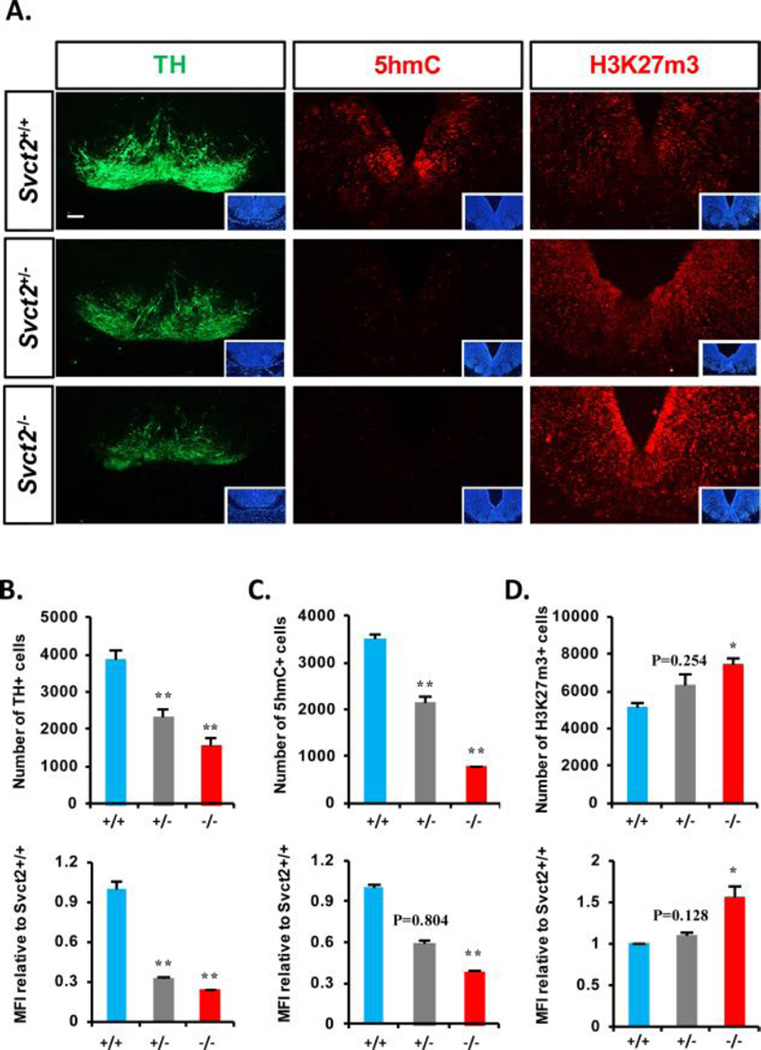
Figure 6. Defect of midbrain dopamine neuron genesis in the midbrain of Svct2 KO embryos.
(A): Representative images of histological immunofluorescent staining of TH (left), 5hmC (middle), and H3K27m3 (right) in E14.5 ventral midbrain (VM) sections of Svct2 wild-type (+/+), heterozygous (+/−), and homozygous (−/−) embryos. Insets are DAPI staining of the section. (B–D): Measurements of TH+ (B), 5hmC+ (C), and H3K27m3+ (D) cell numbers and their individual intensities (MFI) in VM sections are shown from three embryos for each group (derived from two pregnant mothers, three SVCT2+/+, three +/−, two −/− embryos from one litter, and one SVCT2 −/− from the other litter). Statistics are all analyzed in comparison to Svct2+/+. *, p < .05; **, p < .01, n = 3 biological replicates. Abbreviations: MFI, mean fluorescence intensity; TH, tyrosine hydroxylase.

