
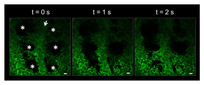
Figure 3. Vacuum enabled thoracic window allows for stable visualization of the pulmonary microcirculation and alveoli in a live C57BL/6 mouse. The pulmonary capillaries surrounding the alveoli (*) can be seen moving in and out of the imaging plane in the z-direction due to the dynamic expansion and contraction of the alveoli (*) with mechanical ventilation. Movement of alveoli starts from the bottom of the image at t = 0 s and moves up to the top with increasing time. Alveoli are marked by asterisks. Intravascular FITC dextran highlights the pulmonary capillaries and a feeding arteriole in green. The open arrow denotes the direction of blood flow within the feeding arteriole. The times displayed are relative to the selected video frames. Scale bars are 20 µm. The feeding arteriole has a diameter of 33 µm, while the capillaries have an average diameter of 6 ± 2 µm. The complete video sequence is included in Movie S1.