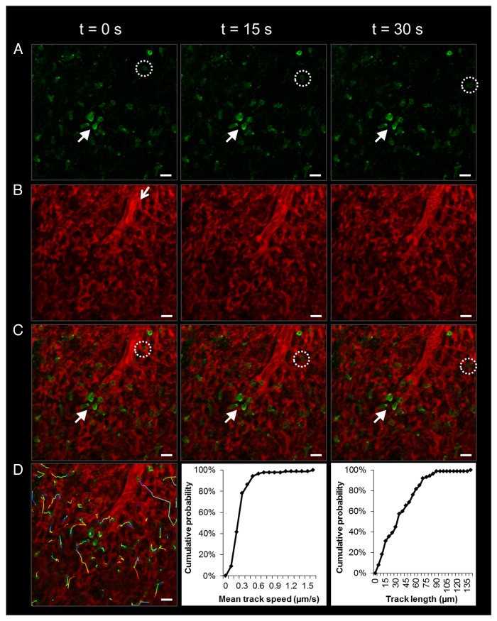
Figure 4. Elongated neutrophils slowly transit or rapidly ‘hop’ through the pulmonary capillaries of a live C57BL/6 mouse. (A) Green channel. Neutrophils (green) were stained by intravascular (iv) injection of FITC Ly-6G mAb. Solid arrow denotes slowly transiting neutrophils. The dotted circle highlights a neutrophil that travels much more rapidly and can be seen hopping between video frames. (B) Red channel. Texas Red-conjugated dextran (red) was administered iv to visualize pulmonary capillaries and a feeding arteriole. The open arrow shows the direction of blood flow within the feeding arteriole. (C) Merged dual color image. Neutrophils (green) are shown trafficking through pulmonary capillaries (red). Neutrophils take on an elongated shape that fills the lumen of pulmonary capillaries. (D) Individual tracks of transiting neutrophils tracked over a 5 min observation period using Imaris software are shown. The tracking video was analyzed in Imaris to extract mean track speed and track length for all the neutrophils and is plotted as cumulative distribution graphs. Note that greater than 90% of neutrophils migrate at 0.5 µm/s or less. The times displayed are relative to the selected video frames. Scale bars are 20 µm. The feeding arteriole has a diameter of 21 µm, while the capillaries have an average diameter of 6 ± 2 µm. The complete video sequences of neutrophil migration within pulmonary capillaries and the tracking video displaying neutrophils with attached dragon tails are included in Movies S2 and S3.

An official website of the United States government
Here's how you know
Official websites use .gov
A
.gov website belongs to an official
government organization in the United States.
Secure .gov websites use HTTPS
A lock (
) or https:// means you've safely
connected to the .gov website. Share sensitive
information only on official, secure websites.
